


Lumbar area osteochondrosis is a chronic degenerative-dystrophic disease that affects the intervertebral disc structure and some lumbar vertebrae located.It affects people who are mostly working.It manifests itself in a variety of symptoms, the main is the pain in the lower back and feet, limiting the movement to the bottom.For diagnostics, research methods such as radiography, computed tomography or lumbar spinal cord resonance tomography are used.In this article, you can be more detailed by the causes, symptoms and methods of diagnosing lumbar spinal osteochondrosis.
Osteochondrosis is the result of body aging.The signs of the disease or other -can be found in almost everyone (!), Starting from 25 years.But here is the severity of this change, their developmental rate, the level of clinical manifestations depends on many reasons, especially how a healthy lifestyle leads to certain people.Moderate physical activity, compulsory morning gymnastics, proper body pose while doing some work (garden, construction, home cleaning etc.), orthopedic mattresses are a time that prevents the development of lumbar spinal osteochondrosis.
According to statistics, spinal osteochondrosis in 80% of cases is the cause of back pain.
How does osteochondrosis develop?
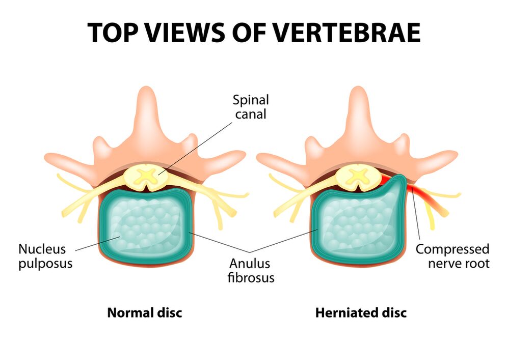
The entire spine consists of a separate vertebra, between the body of the intervertebral disc.That is, between the two vertebrae is a disc.The disc is made up of gelatin nucleus (pulpic) and fibrous rings.The core contains a lot of water and provides depreciation and flexibility of the spinal cord.The fibrous ring is located along the edge of the jacket nucleus, as if holding it in its own.
With the prolonged increase in the jetty core, it changes its physiological properties, loss of water and dry, and finally the sequence: the disc is flattened, and the body of the vertebrae comes close to each other.Along with such a process, in the nucleus jacket, the fibrous ring loses its elasticity and, under the influence of mechanical loads, begins to stand out.This is called protrusion.Then the fibrous rings are cracked, and the gelatin nucleus falls through the resulting gap: the disc hernia occurs.A plot of two adjacent vertebrae and the disc located between them, called the vertebral segment, acquired excessive mobility, thus increasing the load on the nearby segment.The advantages of the neighboring segment trigger the same pathological process in it.This change is called osteochondrosis.
To somehow ensuring spinal stability, bone growth is formed along the edge of the vertebral body, increasing the support area.This phenomenon is called spondylosis.Changes in the joints between the vertebra are called spondylo arthrosis.Usually the third - three pathologies - osteochondrosis, spondylosis, spondil arthrosis - walking nearby.
Cause
Why does osteochondrosis occur?To date, there have been several theories of events:
- Mechanical Theory: Perhaps the main reason should be considered a higher load on the spine.That is why osteochondrosis is an almost obligatory fate of movers, miners, builders and the professions.The occurrence of osteochondrosis of the lumbar area is mainly associated with slopes and lifting severity, forcing uncomfortable work;
- Other factors in development are wrong posture, sitting in the wrong pose, which is very relevant to mental workers;
- Sometimes the role is played by the characteristics of the descendants of the spinal structure and the nutrition of the individual structure;
- Traumatic theory: any trauma to the spine (though most important) can launch the degenerative process;
- Hormone metabolic disorders and endocrine diseases can affect metabolism in the spinal column tissue and contribute to the development of osteochondrosis;
- Age theory implies wearing a natural disc in the process of life.
Rarely, only one of these theories can explain the occurrence of osteochondrosis in each case.Usually at the same time, several factors are "blaming".
In the occurrence of lumbar spinal osteochondrosis, weight gain plays an important role, as it itself is a burden for the spinal column.The higher the body mass index (the degree of obesity), the more obvious change in the spine is usually.Among other causes of osteochondrosis, one can be observed:
- SEDENTARI LIFE;
- Imorly nutrition (fast food, sweeter products, half -finished: all of which lead to trace elements imbalance) and fluid deficiency;
- Spinal structure anomalies (for example, the presence of additional lumbar vertebrae);
- Wear high shoes;
- pregnancy (due to excessive burden on the lumbar spine);
- Sudden termination of training in people who are professionally involved in sports;
- Smoking and alcohol abuse: as a factor that accelerates the aging process in the body.
Symptom
The main manifestation of lumbar spinal osteochondrosis is pain.The nature of the pain, the scene and the direction of the distribution depends on the irritated receptors, that is, how rough changes in the discs and tissues around, there is a highlight or hernia, where the direction of highlights is formed and so on.
Reflex and compression syndrome is distinguished by lumbar spinal osteochondrosis.
Reflex syndrome develops in cases where the receptors of the discs, ligaments and capsules located nearby are irritated.They are reflexive because in addition to pain accompanied by changes in muscle-tonic, vegetative-vascular or neurodistrophic reflexes, that is, irritation with reflexes is transmitted to other structures, causing symptoms mainly from the soft tissue.
Compression syndrome is due to compression (compression) of the nerve roots, blood vessels or spinal cord formed by osteochondrosis by changes.

Lumbar spine reflex syndrome
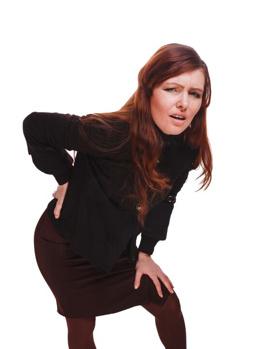
Lumbago(Feelings): pain suddenly - acute pain in the lower back, which occurs with an awkward movement or at the time of physical tension (more often - for no apparent reason).It is believed that the occurrence of lumbago is associated with the movement of the nucleus jacket in the fibrous ring, that is, it develops in the early stages of osteochondrosis.Often the pain is described as a "feeling", "the importance of the lower back."Patients freeze in pose where pain catches them.Little steps cause increased pain (sneezing, coughing, attempting to switch to bed, to move your feet).If a person is in a position that is likely to be in the development of Lumbago (most often), then he or she cannot straighten.The muscle voltage specified in the lumbar spine occurs in reflex.Along the vertebrae in this area, muscle rollers are felt, which can sometimes be seen with the naked eye without touch, and muscle tension is so clear.Feeling painful for the patient.Such increased muscle tone plays a moving role, protecting the affected lumbar segment from pathological mobility, which can cause deterioration in the state.The natural turn of the spinal column at the bottom (lordosis) is flattened, possibly a scoliosis may be due to muscle tension.
Lumbalgia- Another lumbar -level reflex syndrome.This term also means the presence of pain in the lumbar region.But, unlike lumbago, pain does not arise acute, but gradually, within a few hours or even days.Pain is stupid, moderate intensity, increasing during movement, in a sitting or standing position, while moving from one position to another.The slightest release carries a position of lying or back with a roller under the lower back, but the passive increase in the foot straightened in this position causes an increase in pain in the lower back (symptoms of Lassa).Lumbar spine palpation is painful, but muscle reflex tension is less noticeable than with lumbago, and sometimes absent at all.The movement in the lumbar spine is limited, but it is possible.This means that the patient can bend and go to the side to a certain extent (and then the pain increases).
Sciatica- Another various lumbar -level reflex syndrome.This term means the pain in the lower back, which gives the back and the legs (on the back surface).The pain is different, mostly pain, but periodically can be intensified by the type of "fireplace" in the feet.Just like lumbargia, it increases with movement, walking, drowning, reducing behind.Lassa symptoms are usually positive.Lumbar spine palpation hurts, and presses a few points (for example, in the middle of the line separating the back from the thighs, in the middle of the thighs, in the middle of the popliteal fossa).There is a lower back muscle tension.The forward and side -to -side tendencies are limited.
Lumbar spinal compression syndrome
Clinical features depend on which structure is subject to compression.
Among the vertebrae in each intervertebral hole is the nerve root (spinal cord): left and right.If the pathology formation for lumbar spinal osteochondrosis (especially the disc) is rooted, then radiculopathy develops, different symptoms for each root.Common to all radiculopathies in the lumbar region are increased pain during sneezing, cough, lower movement (especially leaning forward), presence of muscle tension in the lower back, restriction of movement in the lumbar spine.The following radiculopathy types of the lumbar spine are the most common:
- Radiculopathy L1, L2, L3: Pain occurs at the bottom, give it to the expected thigh.In the same area, the occurrence of paresthesia (creeping, numbness) is possible, disrupted superficial sensitivity (acute touch from normal, cold and hot feelings) is gone.Low knee reflexes, thigh quadriceps weakened;
- Radiculopathy L4: The pain from the lower back gives to the front of the thigh, the inner surface of the knee joint and slightly lower along the lower surface of the lower leg.In the same area, paresthesia is felt, and the surface sensitivity is lost (reduced).Weakness in the thigh quadriceps also develop, knee reflex decreases;
- Radiculopathy L5: One of the frequent localization.The pain gives the back, along the outer edge of the thigh, along the front surface of the lower leg to the inner edge of the leg and thumb.Paresthesia is felt here, superficial sensitivity is disturbed, and pain is given here during sneezing and coughing.In addition, it is difficult to expand the toes, as the muscles that perform this action are handed over by Kine L5.Sometimes it is difficult to stand on the heel with exposed feet;
- S1 radiculopathy is also often found with lumbar spinal osteochondrosis.The pain gives the back, along the outer edge of the thigh, along the outer edge of the lower leg to the outer edge of the foot and fingers, heels.These zones are characterized by paresthesia feelings, a decrease in surface sensitivity.Achilles reflex is reduced.With damage to this spine, the weakening of the lower leg muscles and the foot flexors develop, so standing and walking in the socks are difficult.
Simultaneous development of radiculopathies some roots is possible, this is a special feature of L5, S1.It happens that one hernia squeezes some roots.
If the herd of the disc returns, it can squeeze the spinal cord.This is only possible when the hernia is located at the top reference point, as there is no spinal cord vertebra under the lumbar vertebra (the spinal cord root is subject to compression, and the horse's tail syndrome develops).
If the vessels in the lumbar region are subject to squash, which runs blood flow to the spinal cord, then in the case of acute circulatory disorders, spinal stroke develops, and with prolonged compression - myelopathy.Myelopathy is characterized by bilateral weakness of the leg muscles, starting from the feet and gradually developing.Sensitivity to the feet is disturbed, the Achilles reflex disappears, and then the knee.It is possible to develop urine disorders (frequently, "important" urgent, which requires immediate satisfaction, urinary incontinence).
Diagnostic method
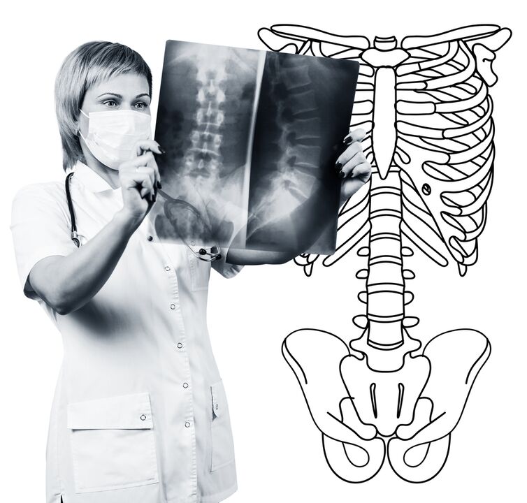
The diagnosis of lumbar spinal osteochondrosis is based on clinical data and additional research method data.The main role belongs to the methods such as:
- Lumbar spine radiographs;
- Tomography is considered lumbar spine;
- Tomography of lumbar spine magnetic resonance.
Lumbar spine radiographs should be done in 2 perpendicular projections and straight side.Such images allow you to see the shape, contour and structure of the vertebral body, the height and shape of the intervertebral disc, the spinal cord, and the natural bend.To display intervertebral joints and intervertebral holes, radiograms are produced in oblique projections.To identify the pathological mobility of the individual lumbar segment (which is a sign of osteochondrosis), radiographs are performed under the terms of the functional trial, that is, in the flexion and extension of the spine.Usually, you can clearly see the height of the intervertebral disc in the front or back in the direction of the body's tendency, with osteochondrosis due to the function block of one segment, the height of the disk does not change either when bending or extending.With pathological mobility, the displacement of the vertebrae forward or backwards is determined.Signs of the main osteochondrosis x include intervertebral gap narrowing, pathological mobility and vertebral body displacement, salt deposition in disc tissue (calcification), regional growth of the vertebral body.Lumbar spinal radiography is a routine of research, which gradually loses its importance to the background of active implementation of new and more informative research methods (CT and MRI).Today's lumbar department radiographs are used as a diagnostic method of inspection.
The lumbar spine CT is also carried out using X -Ray radiation, but the radial load on the body is less than X -Ray.This study was conducted on a special device table - computer tomography, it was really painless.The resulting image is processed using a computer and allows you to see more structures than with spinal radiography.
MRI is a method where electromagnetic radiation is used to create images.The study was also conducted in a position to lie on the table, which calls into the tomography space.MRI is harmless and painless.
The lumbar CT or MRI allows you to see all the spinal structures, carefully examining the intervertebral disc (and jackets and fibrous rings) and intervertebral holes, the contents of the spinal cord.In fact, a small amount of intervertebral disc will not be aware of.This method (especially MRI) allows you to determine the direction of the disc hernia if any, the level of nerve compression, the spinal cord.Therefore, this research method is more informative in the diagnosis of lumbar spinal osteochondrosis than radiography.In addition, they allow you to diagnose not only osteochondrosis, but also other diseases (tumors, blood circulation disorders in the spinal cord, abscesses, congenital defects of the spinal cord and spinal cord), which is important during the diagnosis of differential causes of back pain.
Lumbar spinal osteochondrosis is a disease that most often causes back pain.In fact, the destruction of the intervertebral disc.Due to lumbar spinal osteochondrosis, one often loses work capacity, because, in addition to pain, the disease can cause a violation of spinal mobility, inability to sit, stand and walk.Symptoms of the disease are not specific and require additional research methods to accurately confirm the diagnosis.The most informative and safe diagnosis of osteochondrosis is the spinal MRI.